October 1st, 2021 marks the 50th anniversary of the first computed tomography (CT) scan of a patient, performed at a hospital in the U.K. The half-century is being marked with an article in Physics Today by UC Davis radiologist John Boone and Cynthia McCollough at the Mayo Clinic in Rochester, Minnesota.
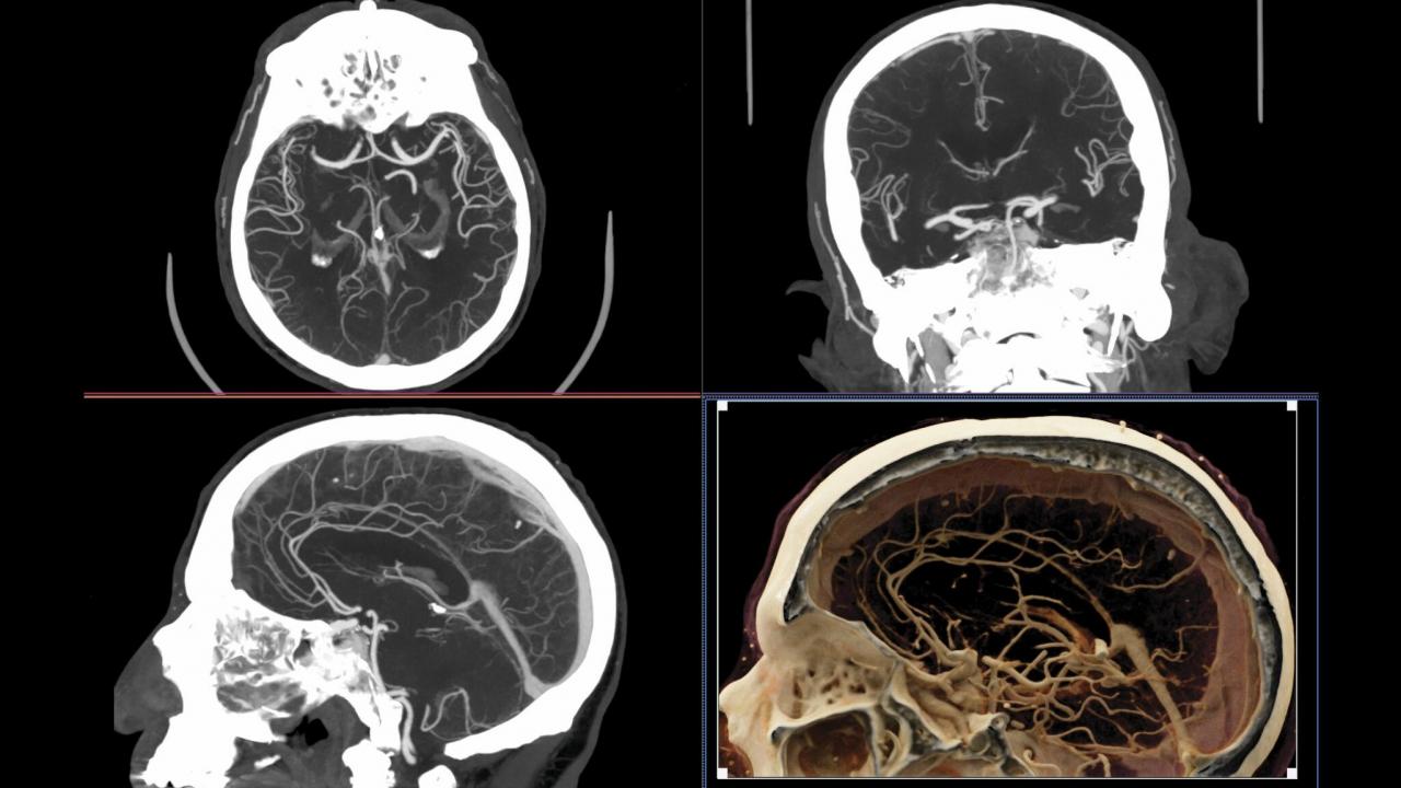
CT scans use thousands of X-ray images to build a three-dimensional image of the body. The technology has revolutionized medicine, allowing physicians to see inside a patient without exploratory surgery. More than 90 million medical CT scans are carried out every year in the U.S.
CT scanning was invented in the research labs of the British music company EMI. Its inventor was electrical engineer Godfrey Hounsfield, who was awarded the 1979 Nobel Prize for Physiology and Medicine along with Allan Cormack, who worked out the mathematics of creating a three-dimensional image from thousands of individual images.
Boone and McCollough describe the principles behind CT scanning and the advances made in the technology in the past 50 years. At first, it was only practical to image the head, as a patient’s head could be immobilized for the several minutes needed to complete a scan. Today, advances in the arrangement of X-ray sources and detectors have made it possible to scan the entire torso from chest to pelvis in five seconds. The most advanced systems can resolve features as small as 150 microns.
CT scans for breast cancer
Boone, a professor of radiology and biomedical engineering and a member of the UC Davis Comprehensive Cancer Center, has himself been an innovator in CT scanning. His laboratory has developed four CT scanners for use in breast cancer patients, using cone beam technology. The technology has been licensed to Izotropic Corporation for commercial development. Boone has also worked with Professors Simon Cherry and Ramsey Badawi on scanners that combines CT and positron emission tomography (PET) for use in breast cancer.
Cherry and Badawi led the team that developed the EXPLORER total-body PET scanner, which has now been developed commercially by United Imaging Healthcare of Shanghai. An EXPLORER scanner is in use at UC Davis Health, and there is also a Mini Explorer for use with small animals at the UC Davis Veterinary Medical Teaching Hospital.
Media Resources
Computed Tomography Turns 50 (Physics Today)